
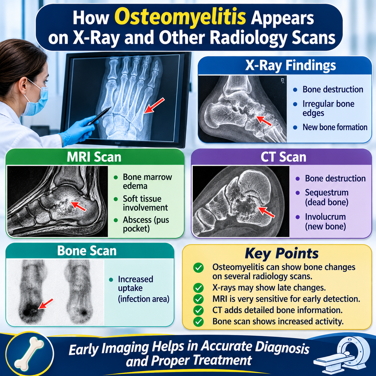
Osteomyelitis is a serious bone infection that requires timely diagnosis and appropriate medical treatment. Imaging examinations help doctors identify bone damage, assess surrounding tissues, and determine how far an infection has spread. Osteomyelitis radiology plays an important role in recognising early abnormalities and monitoring treatment progress.
However, different imaging techniques reveal different stages and features of bone infection. An ordinary X-ray may appear normal during early disease, whereas magnetic resonance imaging (MRI) can detect changes within bone marrow much sooner. Understanding these differences helps patients interpret their reports and discuss appropriate investigations with their healthcare providers.
What Does Osteomyelitis Radiology Reveal?
Osteomyelitis is an infection involving bone and bone marrow. It commonly develops when bacteria enter the bloodstream, spread from nearby infected tissue, or reach bone through an open wound, injury, or surgical procedure.
Radiological examinations help identify several important features:
- Bone marrow inflammation and swelling.
- Loss of normal bone structure.
- Cortical bone destruction.
- Abnormal periosteal reactions.
- Abscesses within or around bone.
- Dead bone fragments and chronic infection.
- Involvement of neighbouring joints and soft tissues.
The appearance depends on infection duration, the affected bone, the responsible microorganism, and the patient’s overall health.
Imaging findings must always be interpreted alongside symptoms, physical examination, blood tests, and microbiological investigations. No single scan independently confirms every case.
How Does Osteomyelitis Look on an X-Ray?
An X-ray is commonly the first imaging examination ordered when a doctor suspects a bone infection. It is widely available, relatively inexpensive, and useful for identifying established bone abnormalities.
However, conventional radiographs have an important limitation: early osteomyelitis may not produce visible changes.
Early X-Ray Findings
During the first several days of infection, an X-ray may appear completely normal. Infection initially affects the bone marrow, which cannot be examined directly in sufficient detail using ordinary radiographs.
As the condition progresses, possible findings include:
- Soft tissue swelling around the affected bone.
- Reduced definition of normal tissue boundaries.
- Localised bone density changes.
- Subtle areas of bone destruction.
- Early periosteal reaction.
Bone abnormalities often become more noticeable after approximately 10–14 days, although timing varies considerably between patients.
Advanced X-Ray Findings
When infection becomes established, radiographs may demonstrate:
Osteolysis: Areas where normal bone tissue has been destroyed, producing irregular or darker regions.
Periosteal reaction: New bone formation along the outer surface of an affected bone.
Cortical destruction: Damage to the dense outer layer of bone.
Osteopenia: Reduced bone density around the infected region.
Sequestrum: A fragment of dead bone separated from healthy surrounding tissue.
These findings can raise suspicion of infection, but other conditions, including tumours and certain inflammatory disorders, may produce similar appearances.
A normal X-ray should therefore never be considered sufficient evidence to exclude early infection.
How Does Osteomyelitis Appear on an MRI Scan?
Magnetic resonance imaging is generally the preferred examination when clinical suspicion remains high despite normal or inconclusive radiographs. MRI provides detailed images of bone marrow, muscles, tendons, joints, and surrounding soft tissues.
It is particularly useful because bone marrow abnormalities can develop before obvious destruction becomes visible on X-rays.
Common MRI Findings
Radiologists look for several characteristic changes:
- Bone marrow oedema: Increased fluid within affected marrow, usually appearing dark on T1-weighted images and bright on fluid-sensitive sequences.
- Reduced T1 signal: Normal fatty marrow is replaced by inflammatory material and fluid.
- Increased STIR signal: Infected areas often demonstrate increased brightness on short tau inversion recovery images.
- Cortical disruption: Infection may damage the outer bone structure.
- Soft tissue inflammation: Nearby muscles and tissues may become swollen.
- Abscess formation: Localised collections of infected fluid may develop within bone or surrounding tissues.
What Does Contrast-Enhanced MRI Show?
Doctors sometimes request MRI examinations using gadolinium-based contrast material.
Contrast enhancement can help distinguish inflamed tissue from non-enhancing areas of dead tissue. It also improves assessment of abscesses, sinus tracts, and surrounding infection.
A central area that does not enhance, surrounded by enhancing inflammatory tissue, may suggest an abscess. However, interpretation requires consideration of the complete imaging examination and clinical findings.
MRI is highly sensitive, but marrow oedema is not exclusive to infection. Trauma, recent surgery, fractures, and inflammatory conditions can produce overlapping abnormalities.
What Does Osteomyelitis Look Like on a CT Scan?
Computed tomography (CT) uses X-rays to create detailed cross-sectional images. It provides excellent visualisation of cortical bone and complex anatomical structures.
Although CT is generally less sensitive than MRI for early bone marrow infection, it offers important information about established bone destruction.
Typical CT findings include:
- Irregular cortical bone surfaces.
- Areas of bone erosion.
- Periosteal new bone formation.
- Small gas collections within bone.
- Dead bone fragments.
- Thickened or remodelled bone.
CT is particularly helpful when doctors need to identify sequestra, evaluate complicated bone anatomy, or plan surgical procedures.
Sequestrum and Involucrum on CT
Chronic infection can cause a piece of bone to lose its blood supply and become separated from living tissue. This fragment is called a sequestrum.
The body may respond by producing new bone around the infected area. This surrounding layer is known as the involucrum.
A cloaca is an opening through which infected material can escape from the affected bone.
Together, these abnormalities are important signs of chronic osteomyelitis. CT often demonstrates them more clearly than conventional radiographs.
Can an Ultrasound Detect Osteomyelitis?
Ultrasound uses sound waves rather than ionising radiation. It cannot directly assess bone marrow or reliably exclude infection inside bone.
Nevertheless, ultrasound can provide useful information about nearby soft tissues.
It may reveal:
- Fluid collections beneath the periosteum.
- Soft tissue swelling.
- Superficial abscesses.
- Joint effusions.
- Inflammation near the bone surface.
Ultrasound can also guide needle aspiration, allowing doctors to collect fluid for laboratory testing.
This is particularly useful when a collection is accessible and requires drainage or microbiological examination.
A normal ultrasound does not rule out osteomyelitis. MRI or another appropriate examination may still be necessary when symptoms and clinical findings suggest bone infection.
What Do Nuclear Medicine Scans Show?
Nuclear medicine examinations help identify areas of increased bone activity or infection-related inflammation.
These techniques involve administering small amounts of radioactive tracers that accumulate in particular tissues.
Three-Phase Bone Scan
A three-phase bone scan evaluates blood flow, soft tissue activity, and bone uptake.
Increased tracer accumulation may indicate infection. However, similar findings can occur after fractures, surgery, and other inflammatory conditions.
Consequently, bone scans are sensitive in many situations but may lack sufficient specificity to establish the diagnosis independently.
White Blood Cell Scintigraphy
White blood cell imaging uses labelled white blood cells to identify areas where inflammatory cells accumulate.
It can be helpful when conventional imaging produces uncertain results, particularly in selected patients with previous surgery or implanted hardware.
PET/CT Imaging
Positron emission tomography combined with CT can identify areas of increased metabolic activity.
It may be considered in complicated cases, chronic infection, or situations where MRI is unsuitable.
The choice of nuclear medicine examination depends on clinical circumstances, available technology, and the presence of previous surgery or metal implants.
How Do Radiologists Distinguish Acute and Chronic Osteomyelitis?
The appearance of bone infection changes as the disease progresses. Radiologists consider the duration of symptoms, bone abnormalities, surrounding tissue involvement, and previous imaging results.
Acute Osteomyelitis
Acute infection develops relatively quickly and primarily affects the bone marrow.
Common imaging characteristics include:
- Bone marrow oedema on MRI.
- Soft tissue inflammation.
- Early periosteal changes.
- Possible fluid collections.
- Subtle or absent abnormalities on initial X-rays.
In early disease, MRI is usually more informative than conventional radiography.
Chronic Osteomyelitis
Chronic infection can develop when treatment is delayed, infection persists, or damaged bone remains infected.
Imaging may demonstrate:
- Sequestrum formation.
- Involucrum development.
- Cortical thickening.
- Persistent bone destruction.
- Sinus tract formation.
- Areas of sclerosis and bone remodelling.
Chronic disease can be particularly challenging because old structural changes may remain visible even after active infection has improved.
What Are the Radiological Signs of Osteomyelitis in the Foot?
Foot osteomyelitis deserves particular attention because it frequently develops in people with diabetes, peripheral neuropathy, poor circulation, or longstanding foot ulcers.
The infection may involve the toes, metatarsal bones, heel bone, or other parts of the foot.
On X-rays, doctors may identify cortical erosion, reduced bone density, periosteal reactions, and destruction near an ulcer.
MRI can show whether infection has entered the bone marrow and whether surrounding tendons, muscles, and soft tissues are affected.
One important diagnostic challenge is distinguishing infection from Charcot neuroarthropathy. Both conditions can cause bone destruction, swelling, and changes in joint structure. MRI interpretation must therefore consider ulcer location, clinical findings, bone distribution, and other evidence of infection.
A specialist may also request a deep tissue sample or bone biopsy when imaging findings remain uncertain.
How Do Doctors Confirm Osteomyelitis After Imaging?
Radiology provides valuable diagnostic information, but doctors generally combine imaging with clinical assessment and laboratory investigations.
Important investigations include:
Complete blood count: A raised white blood cell count may indicate infection, although normal results do not exclude it.
C-reactive protein: CRP measures inflammation and can help assess disease activity and treatment response.
Erythrocyte sedimentation rate: ESR may remain elevated during prolonged inflammation and is particularly useful when interpreted alongside other findings.
Blood cultures: These tests can identify bacteria circulating in the bloodstream.
Bone biopsy: A sample of affected bone can be examined and cultured to identify the responsible microorganism.
A positive bone culture can guide antibiotic selection. Superficial wound swabs are generally less reliable because they may identify organisms living on the skin rather than those causing infection inside the bone.
What Treatments Are Recommended After a Positive Scan?
Treatment depends on the infection’s severity, the affected bone, the responsible organism, and whether dead tissue or an abscess is present.
The main medical approaches include:
- Antibiotics selected according to suspected or confirmed bacteria.
- Surgical drainage of accessible abscesses.
- Removal of infected or dead bone when necessary.
- Wound cleaning and appropriate dressing.
- Management of diabetes and circulation problems.
- Pressure reduction and protection of the affected limb.
Some patients require several weeks of antimicrobial treatment. Others need surgery alongside medication, particularly when infection involves extensive tissue damage.
Follow-up imaging is not always necessary at fixed intervals. Doctors generally assess symptoms, examination findings, inflammatory markers, and clinical progress before deciding whether another scan is appropriate.
Can Natural Approaches Support Recovery?
Patients sometimes explore complementary approaches alongside prescribed treatment. However, no dietary supplement or herbal preparation has been established as a reliable way to eliminate bacterial infection inside bone.
Some people investigate Natural Remedies for Osteomyelitis because they want additional ways to support general wellbeing during recovery. A balanced diet, adequate protein, sufficient sleep, hydration, and appropriate nutritional support can contribute to overall health.
However, these measures cannot replace antibiotics, surgical drainage, or removal of infected tissue when clinically indicated.
People taking medication should also discuss supplements with their doctors because certain ingredients can interact with prescribed medicines or affect bleeding and other health conditions.
What Is the ICD-10 Code for Osteomyelitis of the Right Foot?
ICD-10-CM is a diagnostic classification system used in the United States for medical documentation, billing, and healthcare reporting.
There is no single universal code for every case of right-foot osteomyelitis. The appropriate code depends on whether the infection is acute, subacute, chronic, hematogenous, or associated with a draining sinus.
Examples of relevant codes include:
| ICD-10-CM code | Description | M86.071 | Acute hematogenous osteomyelitis, right ankle and foot | M86.171 | Other acute osteomyelitis, right ankle and foot | M86.271 | Subacute osteomyelitis, right ankle and foot | M86.371 | Chronic multifocal osteomyelitis, right ankle and foot | M86.471 | Chronic osteomyelitis with draining sinus, right ankle and foot | M86.671 | Other chronic osteomyelitis, right ankle and foot |
The right-sided designation is important because ICD-10-CM distinguishes between right, left, and unspecified locations for applicable diagnoses.
A radiology report alone does not establish the correct diagnosis code. The treating clinician’s documentation must support the infection type, location, and relevant clinical details.
Diabetic foot infections may also require additional diabetes-related coding. Coders should review applicable sequencing instructions, exclusion notes, and current annual ICD-10-CM guidelines before assigning codes.
Frequently Asked Questions
1. Can an X-ray detect osteomyelitis in its early stages?
Not always. Early infection may affect bone marrow before visible structural damage develops. MRI is generally more useful when symptoms suggest infection but initial radiographs appear normal.
2. Which scan is most accurate for osteomyelitis?
MRI is generally the most sensitive imaging method for detecting early bone infection. CT is particularly useful for examining bone destruction and dead bone fragments.
3. Does osteomyelitis always show on an MRI?
No. Although MRI is highly sensitive, results can occasionally be inconclusive. Previous surgery, metal implants, trauma, and other inflammatory conditions can complicate interpretation.
4. Can osteomyelitis be seen in a blood test?
Blood tests cannot directly demonstrate infected bone. However, CRP, ESR, white blood cell counts, and blood cultures provide supporting information.
5. How long does osteomyelitis take to appear on an X-ray?
Visible bone changes commonly take around 10–14 days or longer to develop. The timing varies according to infection severity and individual circumstances.
6. Can osteomyelitis return after treatment?
Yes. Persistent infection, inadequate blood supply, retained infected bone, and certain underlying conditions can increase recurrence risk. Further clinical assessment and sometimes additional imaging are necessary.
7. Can lifestyle changes treat bone infection?
Healthy eating, proper wound care, and management of underlying conditions support recovery. However, Natural Treatment for Bone Infection should never be interpreted as a substitute for evidence-based antimicrobial treatment or necessary surgery.
8. Should patients use herbal supplements during recovery?
Patients should consult their healthcare providers before taking supplements. Herbal Care Products are not proven treatments for osteomyelitis and should never replace prescribed medical care.
Conclusion
Radiological examinations are essential for identifying bone infection, evaluating its severity, and guiding appropriate treatment. X-rays provide an initial assessment, MRI detects early marrow abnormalities, CT demonstrates structural damage, and nuclear medicine scans can help investigate selected complicated cases.
Understanding osteomyelitis radiology helps patients recognise why different examinations may be recommended at different stages of infection. However, imaging must always be interpreted alongside clinical findings, laboratory investigations, and microbiological evidence.
Anyone experiencing persistent bone pain, fever, swelling, or an infected wound should seek prompt medical assessment. Early diagnosis and appropriate treatment can reduce complications and improve the chances of recovery.